Table Of Content
- 🧐 Are there any risks associated with 3D ultrasounds for the purpose of seeing baby’s hair?
- 🤱 After seeing hair on a 3D ultrasound, can anything be done to enhance hair growth before birth?
- Factors That Determine Whether A Baby Will Have Hair
- 📷 How can I ensure the best possible 3D ultrasound images of my baby?

Premature babies, also known as pre-term babies, are born before 37 weeks of gestation. These babies are often smaller and less developed than full-term babies, which can make it more difficult to see certain features on a 3D ultrasound, including hair. This technology uses a combination of light and sound waves to create an image of the baby. HDlive 3D ultrasound is a type of 4D ultrasound that provides high-definition images of the baby.
🧐 Are there any risks associated with 3D ultrasounds for the purpose of seeing baby’s hair?
Ultrasound reveals teeth and hair growing inside mother's uterus - Yahoo Sports
Ultrasound reveals teeth and hair growing inside mother's uterus.
Posted: Thu, 03 Feb 2022 08:00:00 GMT [source]
Seeing lots of hair on ultrasound might mean your baby’s growing better and has higher hormone levels. Welcome to the world of baby peeping, where the tools of the trade are getting fancier by the minute! Wondering if that’s really lots of hair on ultrasound or just some rogue pixels?

🤱 After seeing hair on a 3D ultrasound, can anything be done to enhance hair growth before birth?
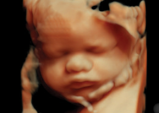
We hope this article has helped explain how ultrasounds show developing hair and how that image compares to the hair your baby will be born with. Although it’s often thought to be old-fashioned, the classic 2D ultrasound may show you the clearest image of your baby’s hair. The hair that a baby is born with generally sheds within the first six months after birth. As we mentioned before, lanugo is a temporary type of hair that developing babies grow.
Factors That Determine Whether A Baby Will Have Hair
As a mother of three young children myself, I have firsthand experience when it comes to walking through difficult situations as a parent. My commitment is to share my own experiences as well as research-based information in order to provide insightful advice for all kinds of issues related to parenthood. Additionally, certain medical conditions can cause babies to be born without hair, such as alopecia or trichotillomania. Once these techniques have been used, technicians may be able to detect slight variations that indicate a baby’s growing locks. The entire process usually takes about 20 to 30 minutes, and you’ll be able to watch the images on a screen as they’re captured.
Baby Born With Lots of Hair on Body
Determining a baby’s gender is one of the most exciting parts of pregnancy for many parents. While there are many old wives’ tales and myths about how to predict the sex of a baby, a 3D ultrasound is one of the most reliable ways to determine the gender of a baby before birth. There are many myths surrounding pregnancy, and two of the most common ones are related to hair and heartburn.
You’re Pregnant! How These Moms Reacted
The Editorial Team is comprised of several freelance hair enthusiasts that share a love of hairstyles, haircare, and hair products. Using both personal experience and third-party research, the team brings a unique perspective to their writing that might even feel like your hairstylist is talking to you themselves. When a patient may have cancer or some other abnormal cell growth, ultrasounds are indispensable when externally examining whatever problem has arisen.
Overall, while it is possible to see hair on a 3D ultrasound, the visibility of hair strands and follicles can vary depending on factors such as hair thickness, color, and growth. Vellus is the hair a baby is born with, usually formed in the last weeks of the third trimester. Predicting your baby’s look can be difficult and sometimes inaccurate because no one baby is born the same. Similarly, if both parents have a lot of hair, the baby may be born with a fuller head of hair.
📷 How can I ensure the best possible 3D ultrasound images of my baby?
It uses advanced technology to produce realistic images that resemble a photograph. Overall, interpreting ultrasound images requires a combination of knowledge and practice. By paying attention to the texture, shadows, contours, and fuzzy halos of ultrasound images, it is possible to gain a better understanding of the structures and tissues that are being imaged. The hair on the scalp of the fetus grows at a rate of about 0.5 mm per day during the third trimester. The hair growth is influenced by the hormones produced by the mother, including estrogen and progesterone. During the first trimester, the baby’s hair starts to grow in the form of fine, soft hair called lanugo.
The second ultrasound, between 18 and 22 weeks, is to check the fetal anatomy for abnormalities, infections, and growth. Issues with fetal anatomy are typically detected at the 20-week anatomy scan if they are present. On a 2D ultrasound, hair strands will appear bright white in contrast to the darker background.
The amount and thickness of hair a baby has at birth are influenced by genetics. A complicated or high-risk pregnancy will often require more frequent ultrasounds during the first and third trimesters. It is normal for some babies to be born without hair either due to genetics or lack of estrogen. From the What to Expect editorial team and Heidi Murkoff, author of What to Expect When You're Expecting. What to Expect follows strict reporting guidelines and uses only credible sources, such as peer-reviewed studies, academic research institutions and highly respected health organizations. Learn how we keep our content accurate and up-to-date by reading our medical review and editorial policy.
3D printed human hair - polymer continuous fiber reinforced composites through Vat Photopolymerization process - ScienceDirect.com
3D printed human hair - polymer continuous fiber reinforced composites through Vat Photopolymerization process.
Posted: Fri, 28 Apr 2023 19:40:21 GMT [source]
However, it is important to use it appropriately and in conjunction with other prenatal tests and screenings to ensure the health and well-being of both the mother and the baby. Although you may see some fuzzy white strands of hair on your baby’s head at around seven months of pregnancy, your baby will likely lose this lanugo before birth. Today's Parent reported that babies usually lose their lanugo between weeks 32 and 36, and that premature babies might be born with their protective fur. In premature babies, lanugo eventually falls out and is replaced by vellus, the "peach fuzz" that grows on hairless areas of the body (feel your earlobe and see for yourself). If you do see hair in a late ultrasound, it will probably look like white strands on the scalp, or a fuzzy white halo.
During pregnancy, 3D ultrasounds can be used to monitor fetal growth and development, detect abnormalities or birth defects, and determine the gender of the baby. In addition, 3D ultrasounds can provide valuable information about fetal cardiac activity and help healthcare providers diagnose and treat any potential issues. Although medically necessary ultrasounds aren’t shown to cause harm when performed by trained health care professionals, ultrasound may heat tissues or produce small bubbles called cavitation. As we discussed earlier, 3D ultrasounds create three-dimensional, lifelike images of your baby in the womb. These detailed images give you a better view of your baby’s facial features, limbs, and other physical characteristics. While 3D ultrasounds are not typically used for routine prenatal care, they can sometimes provide additional information about your baby’s development or be used for keepsake purposes.
The American College of Obstetricians and Gynecologists (ACOG) recommends that 3D ultrasounds should only be performed when there is a medical indication for the procedure. This means that it should only be done when there is a specific medical reason, such as identifying a potential abnormality or monitoring fetal growth. Before the procedure, the patient will need to schedule an appointment with a health care provider who specializes in ultrasound imaging.
Like 3D ultrasounds, 4D ultrasounds are not a standard part of prenatal care, but they can be an exciting way for parents-to-be to bond with their baby before birth. Medical ultrasounds are typically recommended by healthcare providers for diagnostic or monitoring purposes. These ultrasounds are performed by trained professionals who use specialized equipment to obtain images of the fetus.
Due to its small size and light color, fine baby hair may not be visible until later in the pregnancy when it has grown thicker and darker. It’s important to note that while 3D ultrasounds can be a fun and exciting way to connect with your baby before birth, they are not a medical necessity. Traditional 2D ultrasounds are still the go-to for prenatal check-ups and monitoring your baby’s development. Ultimately, the decision to have a 3D ultrasound comes down to personal preference, and it’s always wise to discuss your options with your healthcare provider.